Key points from article :
A holographic system discovers and recognizes disease signs by analyzing tens of thousands of cells per minute.
Distinguished healthy samples and either cancerous or carcinogen-exposed, pre-cancerous cells with nearly 100% accuracy.
Simpler and cheaper screening technology for use in remote, low-resource settings.
"..points toward the potential for point-of-care diagnostics," - Adam Wax, co-author of the study.
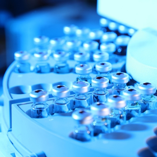
Sample is passed beneath a camera, which can determine the topography and features of each cell.
"It takes about 30 seconds to process a 1ml sample of over half-million cells," - Cindy Chen, first author.
Categorizes each cell using the 2D area it occupies, the 3D space it takes up, its shape and average height.
Measurements are combined with data points and deep learning techniques to spot the disease.
"A pathologist could pull up individual data for any of the cells that were imaged for closer inspection," - Chen.
Study by Duke University published in the journal Frontiers in Physics.