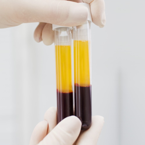
The search for ways to slow down or reverse aging has fascinated humans for centuries. From ancient myths to modern science, the idea of rejuvenation has always captured our imagination. Recent studies on heterochronic parabiosis (HCPB) have provided new insights into how young blood can influence aging.
Researchers have found that young blood can rejuvenate old tissues, while old blood can induce aging in young tissues. But what are the specific factors in young blood that drive these changes?
Unveiling the Role of PEDF
In a groundbreaking study, scientists identified pigment epithelium-derived factor (PEDF) as a key player in the rejuvenating effects of young blood. This secreted protein has emerged as a systemic mediator of rejuvenation. Researchers used an innovative in vitro HCPB system to show that PEDF can extend the replicative lifespan (RLS) of human primary fibroblasts.
This discovery highlights the significant role of PEDF in maintaining cellular health and delaying aging. Understanding the mechanisms and effects of PEDF could lead to new therapeutic approaches for age-related conditions and enhance overall health and longevity.
The In Vitro HCPB System: A New Frontier
Traditional HCPB studies involve surgically connecting the circulatory systems of young and old animals. While effective, this method is time-consuming and resource-intensive.
To overcome these challenges, researchers developed an in vitro HCPB system using human fibroblasts. This system allows for high-throughput screening of circulating factors, making it easier to identify those responsible for aging and rejuvenation.
The Impact of Serum Age on Cellular Health
The study began by examining how the age of serum affects the health and lifespan of human fibroblasts. Researchers cultured fibroblasts in serum from donors of various ages, including fetal bovine serum, and serum from donors aged 1.5, 3, 6, and 10 years.
They found a clear inverse relationship between the age of the serum donor and the lifespan of the fibroblasts. Specifically, fibroblasts cultured in older serum exhibited reduced replicative lifespan (RLS) and increased signs of cellular aging.
These signs included higher levels of senescence markers, such as p16INK4A expression, loss of LaminB1, increased senescence-associated β-galactosidase activity, and a lower percentage of actively replicating cells indicated by Ki67 positivity.
This reduction in cellular health with older serum was confirmed through RNA-seq analysis, which showed significant changes in gene expression related to cell cycle regulation, inflammation, and stress responses. The findings indicated that the components present in aged serum directly and adversely affect cellular function and longevity.
This foundational work set the stage for identifying specific factors within young serum that could counteract these negative effects, leading to the discovery of PEDF as a rejuvenating factor capable of extending the lifespan of human fibroblasts when added to aged serum. This research highlights the potential of targeting serum components to combat aging and improve cellular health.
PEDF in mice
Through their in vitro HCPB system, the researchers identified PEDF as a key factor that declines with age in mammals. They discovered that supplementing aged serum with PEDF significantly extended the RLS of human fibroblasts. This finding suggests that PEDF plays a crucial role in maintaining cellular health and delaying the onset of aging.
To validate their findings, the researchers administered PEDF to aged mice. The results were remarkable. PEDF treatment improved cognitive function, reduced hepatic fibrosis, and decreased renal lipid accumulation in the mice. These improvements highlight the potential of PEDF as a therapeutic agent for combating age-related decline.
Mechanisms of Action
The study delved into the mechanisms through which PEDF exerts its rejuvenating effects on cells. Using RNA-seq analysis, researchers investigated the changes in gene expression when aged serum was supplemented with PEDF. The results were striking: PEDF supplementation significantly countered the adverse impacts of aged serum on cellular gene expression profiles.
Specifically, PEDF downregulated the expression of genes associated with cell stress, inflammation, and senescence. These genes are typically involved in pathways that lead to cellular aging and dysfunction, indicating that PEDF helps mitigate the harmful effects associated with aging.
Conversely, PEDF upregulated genes involved in crucial cellular processes such as cell cycle regulation and mitosis. These processes are essential for maintaining cell health and ensuring proper cell division and replication. By enhancing the expression of these genes, PEDF promotes a more youthful and functional cellular environment. This dual action of reducing harmful gene activity while boosting beneficial processes underscores PEDF's role as a potent mediator of cellular rejuvenation.
The findings suggest that PEDF helps restore a more balanced and healthy gene expression profile in aged cells, promoting longevity and improved function. This mechanistic insight into PEDF's action opens new avenues for developing therapies aimed at combating age-related decline and enhancing tissue health through targeted gene expression modulation.
Broader Implications and Future Directions
The identification of PEDF as a systemic mediator of rejuvenation opens up exciting possibilities for aging research. It suggests that targeting specific circulating factors could delay aging and improve overall health. Furthermore, the in vitro HCPB system provides a valuable tool for screening other potential geroprotective factors.
The discovery of PEDF and its rejuvenating effects marks a significant step forward in our understanding of aging. By unlocking the secrets of young blood, scientists are paving the way for new therapies that could enhance health and extend lifespan. As research continues, we move closer to realizing the dream of slowing down, or even reversing, the aging process.
The study was carried out at Albert Einstein College of Medicine and published in bioRxiv.


