Join the club for FREE to access the whole archive and other member benefits.

Microbubbles provide clear images of blood vessels in live animals

25-Nov-2015

Key points from article :

Technique takes advantage of how high-frequency ultrasound waves interact with tiny gas-filled bubbles about 2 micrometres in diameter.

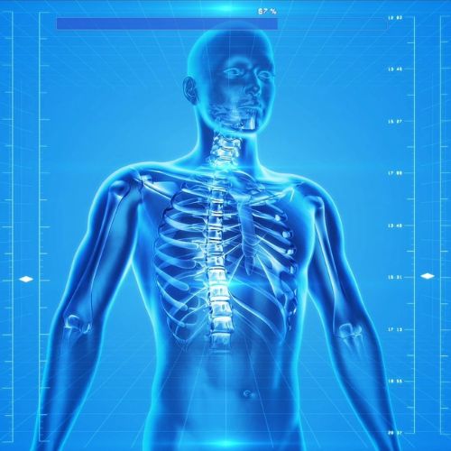
Allows doctors to image organs deep in the body in microscopic detail – pixels about the size of a red blood cell.

Other techniques make compromise between clarity, speed and organ depth.

Could help understand diseases that modify blood-vessel systems, e.g. diagnose what stage a cancer is at and better understanding of stroke.

The study was published in the Nature journal.

Mentioned in this article:

Click on resource name for more details.

Langevin Institute

Academic research institute focusing on wave phenomena and imaging.

Mickael Tanter

Research Professor at INSERM, Member of the European Academy of Science, Co-Founder of Supersonic Imagine, CardiaWave & Iconeus companies.

Nature

Scientific journal covering research from a variety of academic disciplines, mostly in science and technology

Topics mentioned on this page:
Diagnostics