Key points from article :
Microscopes are effectively 2D imaging devices that only detect light, can’t sense clinically important properties.
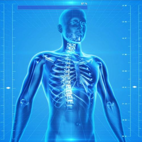
Ultrasonic imaging can provide nuance about the 3D structure.
Also, waves can offer a lot of information about the nature of the tissues themselves.
“... system’s ability to measure the stiffness of a specimen, its bio-compatibility, and its endoscopic-potential ... set it apart. ... the ultimate goal of minimally invasive point-of-care diagnostics,” said Dr Salvatore La Cavera III, team leader.
The tiny size of the new probe allows it to be used alongside optical components of conventional endoscopes.
It can be used to visualize difficult to reach targets such as those within the GI tract.
Uses two lasers that emit short pulses of energy to stimulate and detect vibrations in a specimen.
By detecting these “collided” laser pulses, the shape of the traveling sound wave can be recreated and displayed visually.
Research by University of Nottingham published in Light: Science & Applications.